Our Research & Expertise
Chronic Kidney Disease
We are studying the mechanism of kidney injury and repair with the ultimative goal to find new treatments for the vast and growing patient population suffering from chronic kidney disease (CKD).
Heart Failure
Chronic heart failure is the leading hospital discharge diagnose and the killer of patients with chronic kidney disease. We are studying key signaling pathways between pericytes, cardiomyocytes and endothelial cells to develop novel therapeutics in heart failure and uremic cardiomyopathy.
Fibrosis
Fibrosis has been estimated to be involved in almost 50% of all deaths in the developed world. We are studying fibrosis of heart, kidney, lung, vasculature and bone-marrow with the goal to develop novel targeted therapeutics.
Vascular Disease
We are performing state of the art genetic fate tracing and omics research to understand the key cellular and molecular events in athero- and arteriosclerosis with a special focus on vulnerable patients with chronic kidney disease and / or diabetes.
CRISPR/Cas9 gene editing
We use in vitro and in vivo CRISPR/Cas9 gene editing to validate pathways and therapeutic targets.
3D cell-culture modeling & Stem Cells
We are working with 3D cell culture models of human and mouse cells for disease modeling, target validation and compound screening.
In vivo studies & translation
We are using various transgenic mouse models for disease modeling and genetic fate tracing and validate findings in human tissue and serum.
Genomics & Proteomics
We are performing various single cell genomic technologies and proteomics to understand cell-fate in disease, heterogeneity and cross-talk with the ultimate goal to identify novel biomarkers and therapeutic targets.
OUR TEAM

Rafael Kramann, MD, PhD, FASN

Jana Lippe

Sikander Hayat, PhD

Judith Sluimer, PhD

Marian Clahsen van Groningen, MD, PhD

Claudia van Roeyen, PhD, Priv. Doz.

Turgay Saritas, MD, Priv. Doz.

Sylvia Menzel, MSc, AMRSB

Nadine Kaesler, PhD

Susanne Ziegler, PhD
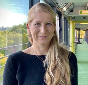
Anne Babler, PhD

Konrad Hoeft, MD

Tore Bleckwehl, PhD

Katharina Reimer, MD

Jitske Jansen, PhD

David Schumacher, MD

Vivien Goepp, PhD

Noriyuki Yamashita, MD, PhD

Xiaohang Shao, MD

Veronica Herranz, PhD

Isaac Shaw, PhD

Felix Schreibing, MD

Ling Zhang, MD

Marius Kohl, MD

Lars Koch , MD

Yuchen Liu, MD

Maurice Halder, PhD

Xinyu Wu, MD

Gideon Schaefer, MD

Qinqing Long, MD

Peggy Jirak

Imma-Katrin Härthe

Larissa Tenten

Nina Graff

Ursula Förster

Isabelle Badziong

Liliane Palm

Sadaf Ijaz, MSc

Max van der Velde

Sidrah Maryam, MSC

Daphne Bouwens, MSC

Lea Ade

Carla Schikarski

Ritabrata Sanyal

Anne-Sophie Andries

Suchanda Bhattacharyya

Christian Möller

Fabian Schelke

Anna Mueller
Join our Team
Recent Work
selected publications from the lab
If you are interested in supporting our research by donation and help to develop novel therapeutics for the vast and growing population of patients suffering from organ fibrosis, heart failure and chronic kidney disease you can contact Dr. Kramann via email
LATEST NEWS FROM THE LAB
ERC Funding for Kramann Lab
Read full article

